


The main source of non-natural exposure
Editorial revision 2026
Apart from natural radioactivity, the main source of exposure to radiation comes from medical diagnostics and therapies. Exposure to radiation for diagnostic purposes varies enormously depending on a person’s state of health, from 0 for someone who does not see a doctor to several tens of millisieverts (mSv) for someone who has to undergo several CT scans.
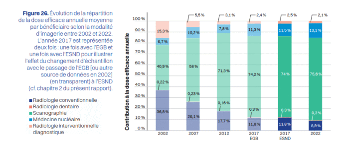
Distribution of diagnostic procedures and collective effective dose by imaging modality. Source: Michel, C., Sage, J., & Isambert, A. (2025). Population exposure to ionising radiation from diagnostic medical imaging procedures performed in France in 2022 (ExPRI expert report). French Nuclear Safety and Radiation Protection Authority (ASNR). Available on the HAL-ASNR portal.
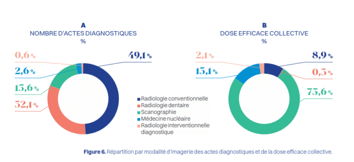
Contribution of different medical examinations to the dose
Conventional and dental radiology account for by far the largest
number of procedures performed. CT examinations can be seen to
contribute substantially to the effective dose for the population.
Source: Michel, C., Sage, J., & Isambert, A. (2025). Population exposure to ionising radiation from diagnostic medical imaging procedures performed in France in 2022 (ExPRI expert report). French Nuclear Safety and Radiation Protection Authority (ASNR). Available on the HAL-ASNR portal.
Radiation exposure in medicine is due much more to the use of X-rays for radiography than to radioactivity itself. It is more accurate to speak of radiation than of radioactivity, even though the radioactivity symbol is displayed in radiology practices. This is a regulatory requirement. But for both the patient and the doctor, it is the effect of the radiation, rather than its origin, that matters.
In diagnostics, the X-rays used are produced by a radiation generator. The rays from a CT scanner come from a small accelerator, which allows them to be directed more precisely. In nuclear diagnostic procedures, such as scintigraphy, gamma rays are of radioactive origin. But whether X-rays or gamma rays, the detection of these rays is similar.
In therapies – radiotherapy or nuclear therapies – the effects of ionising radiation are used. All radiation, regardless of its origin, ultimately deposits its energy by “ionising” living matter. Radiotherapists use X-rays or electrons of a few MeV produced by an accelerator. In nuclear therapy, radiation may be of radioactive origin, as in brachytherapy, or supplied by an accelerator. There are specific applications such as the use of protons (proton therapy).
Targeted radionuclide therapy (TRT) can also be performed. A significant amount of activity is injected into the patient to treat tumours as closely as possible. Radioactive iodine-131 is used for thyroid treatments. Today, new radionuclides are emerging. These include lutetium-177 and short-lived alpha emitters, which are expected to undergo major development (Pb-212, Bi-213, Ac-225, Ra-223, Ra-224, etc.).
No one would think of questioning the advances in medicine that have led to increased life expectancy, advances due, to a large extent, to the radiation exposure required for X-rays, from simple radiographs to CT scans, and to today’s nuclear medicine. Over the years, the development of increasingly sensitive detectors has made it possible to reduce radiation exposure while achieving the same quality of results.
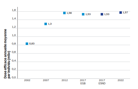
Evolution of the average annual effective dose per beneficiary between 2002 and 2022. In 2002, the dose was 0.8 mSv per year per inhabitant (on average). Since 2012, this value has doubled (1.6 mSv) and corresponds to approximately 25 to 30% of the population’s total exposure. Source: Michel, C., Sage, J., & Isambert, A. (2025). Population exposure to ionising radiation from diagnostic medical imaging procedures performed in France in 2022 (ExPRI expert report). French Nuclear Safety and Radiation Protection Authority (ASNR). Available on the HAL-ASNR portal.
Doses can also be broken down according to the sex of the population.
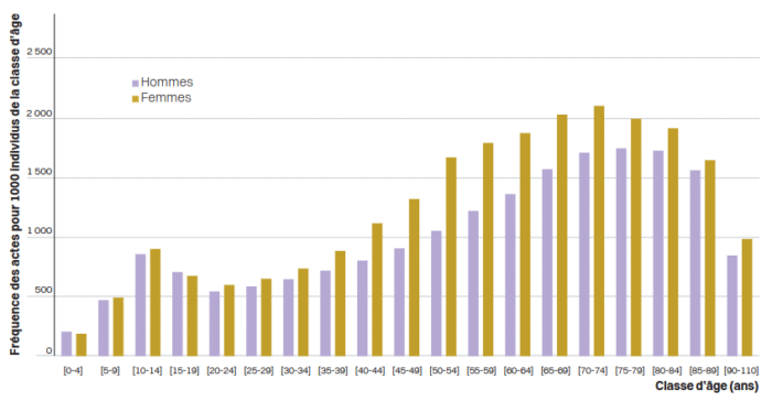
Frequency of procedures (all modalities) according to age group and sex (expressed as number of procedures per 1,000 beneficiaries). Source: Michel, C., Sage, J., & Isambert, A. (2025). Population exposure to ionising radiation from diagnostic medical imaging procedures performed in France in 2022 (ExPRI expert report). French Nuclear Safety and Radiation Protection Authority (ASNR). Available on the HAL-ASNR portal.
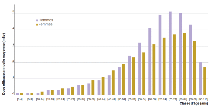
Average annual effective dose per beneficiary according to age group and sex. It can be seen that women undergo more procedures. This corresponds, in particular, to national screening campaigns for breast cancer and osteoporosis. However, in terms of dose, men are more exposed. Mammography and bone densitometry are low-dose examinations, unlike prostate cancer treatment, for example.
Source: Michel, C., Sage, J., & Isambert, A. (2025). Population exposure to ionising radiation from diagnostic medical imaging procedures performed in France in 2022 (ExPRI expert report). French Nuclear Safety and Radiation Protection Authority (ASNR). Available on the HAL-ASNR portal.