Exposures to radiations in diagnostic radiology
The vast majority of diagnoses based on radiation (X-rays, fluoroscopy, scanners) use X-rays. These rays usually coming from the deepest layers of the atom do not result from nuclei and have nothing to do with radioactivity. But when health is at stake, the source of emission, relevant for the physicist, becomes secondary. It is the dose that matters to the patient. The doctors prefer to speak of ionizing radiation. These pages would be incomplete without information on doses from X ray examinations
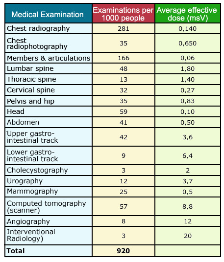
X-ray doses
(Adapted from UNSCEAR 2000.)
© UNSCEAR
On average, the number of these examinations is approximately 1 per year per capita in developed countries. Examinations by far the most common are radiographs of the chest and limbs and joints, but the doses are low or moderate.
In the hierarchy of doses, classical radiographs like pictures of the chest are the less irradiating: a fraction of millisievert (mSv). Some radiation protectionists estimate the risk of dental radiography – 0.020 mSv – equivalent to that of smoking a single cigarette. The radiographs of the spine, pelvis, and digestive system are more irradiating.
At the top of the hierarchy are scanners that are X-ray examinations at multiple angles, angiography and interventional procedures in which the intervention of the surgeon is guided by an x-ray camera.
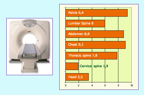
Typical doses in Computed tomographies
(Source: KGGerber, adapted from UNSCEAR 2000)
© IN2P3
As in nuclear medicine, an extensive attention is devoted to radiation protection aspects, which allowed to greatly reduce the doses received by patients and by medical staff (remember diseases of the first radiologists who were exposed to radiation without knowing its dangers).
More specific precautions should be taken in the following cases:
– In pediatrics: Due to the high sensitivity of the organism during growth period.
– Scanner examinations: Because of relatively high doses and the character more and more frequent of these examinations.
– Angiography (X-ray of the venous system): Due to relatively high doses for the patient.
– Interventional Radiology: Due to the relatively high exposure to the patient and the physician.
– Radiography of the end of the large intestine and, to a lesser extent, lumbar spine, pelvis and hip: Due to the proximity of the genitals and the possibility of genetic effects.
–Screening examinations: Mammograms, lung, and stomach because of the large number of people involved.
This animation by UK not-for-profit Nuffield Health shows just how little radiation medical scans expose us to by comparing the levels to natural background radiation, air travel and even space travel. Produced with the help of the Radiological Protection Centre in London.
Other articles on the subject « Expositions in Medicine »
Radiation doses in diagnostics
From simple radiology to PET scans and scintigraphy Scintigraphy scans are by far the most common[...]
Doses (nuclear diagnostics)
Nuclear medicine : doses in nuclear diagnostics Nuclear medicine examinations are performed only [...]
Doses with therapies
Radio and nuclear therapies: Doses are high but local Genuine nuclear therapies (brachytherapy, p[...]
Doses in Nuclear therapies
High local doses In nuclear therapy, the doses delivered to tumours amount to few tens of Grays. [...]