PET : an instrument for early cancers detections
Positron emission tomography, or PET, is a technique of Nuclear Imaging. In full expansion, usually associated with a scanner, it allows the early detection of cancers.
Positron emission tomography (PET) grants today medical teams the ability to carry out accurate diagnoses of patients before a disease has had time to spread, as well as to monitor recoveries from surgical operations. In the field of cancer treatments, PET scans allow oncologists to see how patients cope with radiotherapy or chemotherapy, and help in the identification of any potential tumours. A diagnosis obtained with a PET scan can change the way doctors follow a patient, by stopping inefficient treatments, replacing a planned surgery with radiotherapy, or even changing the nature of an intended surgery or therapy.
Radioactive tracers used are beta-plus radioelements, emitters of positive electrons or positrons. After a short range, positrons vanish emitting two back to back gamma rays. The simultaneous detection of this gamma pair allows to locate the zone of emission that is close to the fixation of the radioactive tracer. With many pairs detected, physicists can draw the map of where the radioactive atoms have been fixed by our cells in order to screen the hot spots.
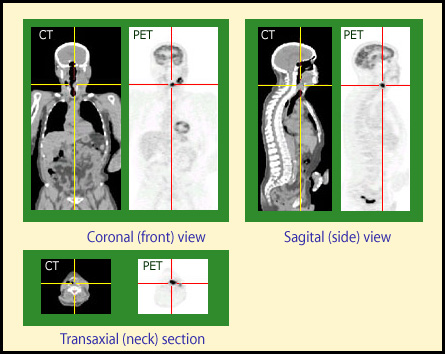
PET Images
PET procedures allow for the early discovery of cancers and are useful in following the evolution of cancerous growth. These three images of the same patient taken with a CT (Computed Tomography) scanner and a PET camera show the advantages of nuclear imaging techniques. While the scanner provides detailed anatomical images but is unable to detect any lesions, the PET images show an abnormal cellular activity in the larynx close to the principal tumour. Tomography techniques allow for pbtain three body views simultaneously.
© General Electric/Medical systems (University of Zurich courtesy)
As opposed to iodine 131 or technetium 99m, which are isotopes commonly used in scintigraphy, positrons emitters are either light chemicals abundant in the human body (such as oxygen, carbon, nitrogen etc.) or halogens (fluorine 18 or bromine 76) which can easily be incorporated to other molecules. This means that chemicals which play a key role in the human metabolism can be marked with radioactive elements and thus followed around the body. The way human tissue absorbs glucose, for instance, can be mapped after an injection of fluorodeoxyglose (FDG), a chemical marked with radioactive fluorine 18. The absorption of glucose is an important parameter in monitoring a person’s health, as it reveals key information about the way different tissues work – whether in the brain, the muscles of the heart or cancerous tumours.
A molecule as simple as water, once it is marked with oxygen 15, can be used to follow the way blood flows through the brain and thus provide a greater understanding of the way it works. These explorations have helped pave the way for new insights in neurology, psychology, linguistics and the cognitive sciences.
Unfortunately, beta-positive isotopes do not exist in nature and must be created in the laboratory.
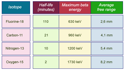
Characteristics of positron emitters:
One can see from the above table that radioisotopes used in tomography have particularly short half-lives. Some are so brief that the products must be used in the same place as their manufacture, with only fluorine 18 lasting long enough for transportation to be worthwhile. The gamma photons which form the image are created after the emitted positrons travel a few millimetres in the body. The shorter the distance travelled, the more accurate the final image.
© IN2P3
The half-lives of these artificial positron emitters are very short: oxygen 15 has a two minutes half-life while the amount of fluorine 18 halves in just under two hours. These radioisotopes must therefore be inserted into the body as soon as possible after they are created, and so many hospitals have a cyclotron (a mini-accelerator) to produce these isotopes and a radiochemical laboratory to extract them.
Of all the molecules marked with positron emitters, FDG is by far the most important. Apart from its remarkable biological properties, its ‘comparatively long’ half-life means that it can be transported from its production site to numerous hospitals in the area. The most common use of FDG in PET scans performed today is the full-body scan performed in clinical oncology to determine the spread of a cancer.
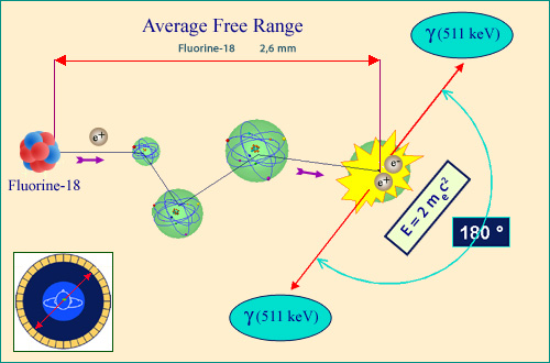
Two gamma rays emitted back-to-back
Positrons emitted by a beta-plus marker disappear after travelling a few millimetres through the body, where they annihilate with an atomic electron in a process whereby both particles cease to exist. The 2×511,000 electronvolts released are carried away by two gamma photons emitted back-to-back. In PET, the travel paths of both photons are traced back to the source point – a distance which is usually comparatively small. Fluorine 18, with its comparatively long half-life, is a frequently-used marker
© IN2P3
As of the end of 2002, there were over 530 PET cameras in the United States and 215 in Europe, of which only 11 were in France. A recent program to equip French hospitals with PET scanners has allowed France to make up its deficit since 2003. In 2009, 77 TEP were installed and 28 were in the process to be.
NEXT : PET : principles
NEXT : PET Scan
Other articles on the subject « Nuclear Imaging »
Gamma Cameras
The most widespread of nuclear medical diagnostics Gamma cameras or scintillation cameras are pie[...]
Gamma-camera : principles
Detect gamma rays and reconstruct their line of flight The emission of a single gamma ray is a ve[...]
PET : Principles
Simultaneous detection of two annihilation gamma photons Positron Emission Tomography is an imagi[...]
PET Scan
Fruitful combination of two medical imaging techniques One of the latest technological advances i[...]
Nuclear Imaging History
From radioactive indicators to CT, scintigraphy and PET … The name of Georg de Hevesy is as[...]