Radioactivity, Radiation and Health
The discovery of artificial radioactivity in 1934 gave scientists the ability to create radioactive isotopes of any element they wanted. One of the most important applications of this technology was the development of radioactive tracers, which have helped to revolutionize biology. Using radioactive tracers, it is now possible to understand the way atoms and molecules move through our body: an advance which has led to a better understanding of the human metabolism, and provided an excellent way to verify the effect of drugs and medication.
The exploitation of radioactivity has also led to the development of most important tools in the medical arsenal. Positron Emission Tomography (also known as PET) gives doctors the ability to film the inner workings of the brain, and to detect cancers in the early stages of development. Such techniques, still expensive, remain relatively rare. More widespread is the process of ‘scintigraphy’ – the imaging of gamma radiation with ‘gamma cameras’. Scintigraphy scans can be conducted on bones, the thyroid gland, kidneys, lungs, or even on the muscles of the heart.
The risks patients face from such radioactive scans are minimal when compared to the benefits such tests can offer. It only takes a very small amount of radioactive matter to obtain an accurate scan; most of which have a half-life of a few hours, and disappear from the body quite rapidly. The radiation dose from nuclear medicine diagnostics is approximately the same as that obtained from an X-ray scan: comparatively harmless, but only to be undertaken if the circumstances warrant.
Radioactive isotopes are also used in therapies for the treatment of certain cancers, such as those of the thyroid or the prostate.
Much more often, medicine uses for medical diagnostics and radiotherapies, other sources of radiation than radiation coming from radioactivity : from X-rays of simple radiographies to the gamma rays provided by small accelerators used to perform scanners or treat cancers in radiotherapies.
The treatment of cancerous tumours requires far stronger doses of radiation than for diagnostics. Recent developments have made it possible to treat ever more specific areas of the body, allowing doctors to cut down on the amount of exposure that healthy cells need to experience. A variety of rays are used for this sort of treatment, depending on the end result desired. Whatever technique is used, when such high doses of radiation are involved it is particularly important to choose the right level of exposure, and to follow the guidelines of radioprotection.
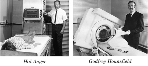
Medical imaging
Two pioneers of medical imaging, Hal Anger and Godfrey Hounsfield inventors of nuclear scintigraphes and CT scanners.
© IN2P3
ACCESS TO CHAPTERS : SUMMARY
– Nuclear medicine : Medical tracers (radiopharmaceuticals), scintigraphic examinations, exposures in nuclear diagnostics, biological analyses.
– Nuclear imaging : Nuclear imaging techniques – Gamma-cameras, Positron Emission Tomography (PET), PET scans
– Radiation therapies : X-ray therapies, nuclear therapies, exposures due to therapies, radiation risks