Simultaneous detection of two annihilation gamma photons
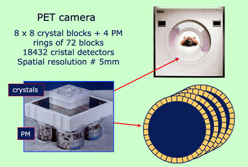
Components of a PET camera
The figure shows the components of a PET camera. One finds at the elementary level, 8 x 8 crystals scintillators viewed by 4 photomultipliers, the whole forming a detection block. 72 of these blocks are assembled into a ring. The final set-up, made of 4 rings, surrounds the bed on which the patient lies. It can move along the axis of the bed. Thanks to these 4 rings, measurements will be taken simultaneously from various angles. This allows the reconstruction of 3D maps of radioactive tracers in the body, while reducing the exposure to radiations. The spatial resolution, ie the precision of reconstruction, is about 5 mm.
© A.Aurengo/ Hôpital Pitié-Salpêtrière (Image Siemens)
Positron Emission Tomography is an imaging technique which maps the distribution of beta-positive emitters throughout the body. The positrons (positive electrons) emitted are identified by the fact that, once they have lost their energy (their range does not exceed a few millimeters), they annihilate with an electron to yield two gamma rays each of 511 keV of energy, emitted back to back. Both gamma reach simultaneously a pair of opposing detectors placed on either side of the annihilation location. Electronic circuits associating these pairs of detectors are designed to identify the annihilation photons.
The two photons are considered relevant if their energies are around 511 keV and their detection times differ by less than one ten-billionth of a second. The collimation is ‘electronic’ as there is no need for lead collimators such as the ones which are used in gamma cameras (although some 2D imaging methods do require such lead collimators).
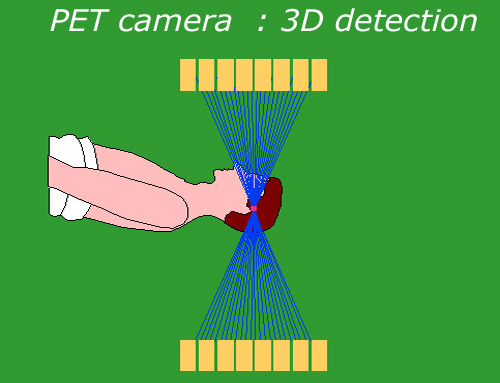
PET camera in 3D Mode
This three dimensional mode is the most common mode of operation of a PET camera. Sensitive scintillator crystals detectors and the coincidence technique remove the need of lead collimators (septa) that were formerly required to select the photons originating from the organ examined. The fact that the direction of the photons is no more imposed by septa increases the sensitivity of the diagnostic and reduce its duration.
© A.Aurengo/ Hôpital Pitié-Salpêtrière
Although the selection in time and the definition of the energy and direction of the gamma are different, the mapping of the spatial distribution of radioactive tracers in the body follows the same principles as in the case of the gamma cameras based on the emission of single gamma photons.
The complex mathematical algorithm is due to Godfrey Hounsfield and Allan Cormack who received the 1979 Physiology or Medicine Nobel Prize for their theory of « Computed Tomography » Combining data obtained from different angles allows for the construction of a series of tomographic cross-sections of the patient’s body. These planar projections can then be used to three-dimensional image of where the radioactive tracers are located in the patient.
Unlike the case of gammas camera, various types of PET camera are available. The scintillating crystal can be made of sodium iodide NaI (Tl), Bismuth germanate (BGO), lutetium oxyorthosilicate (LSO) or even gadolinium oxyorthosilicate (GSO).
It is also possible to use a two-headed gamma camera to detect coincidence events, by increasing the thickness of the NaI (T) crystals and adding an electronic system capable of registering such coincidence events. This technique is known as Emission Tomography with Coincidence Detection (ETCD).
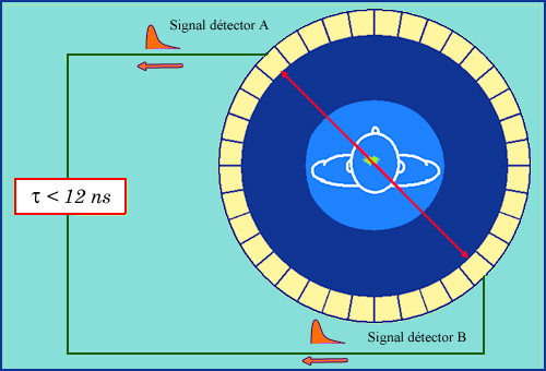
Coïncidence circuit
The two gamma rays emitted back to back during the positron annihilation are detected simultaneously by two opposite scintillators. This coincidence is a very strong signature that distinguishes them from other photons. Specific electronic « coincidence » circuits pick up gamma pairs. On the figure, it is requested that the signals coming from the scintillators A and B coincide within 12 billionths of a second (nanosecond). The straight line joining the centers of detectors A and B is an approximation of the actual line of flight of the two gamma rays.
A.Aurengo/ Hôpital Pitié-Salpêtrière
Cameras designed for scans involving positron emitters have all an annular (ring-like) structure. Scans with cameras of this kind are generally faster and offer much higher-resolution images than those with ETCD cameras.
Finally, the most recent PET scanners can have a camera attached to them (PET/TDM cameras), resulting in a hybrid apparatus which takes anatomic images from the scanner and superimposes them on the functional images from the PET module.
NEXT : PET Scan
RETURN : Positron Electron Tomography
Other articles on the subject « Nuclear Imaging »
Gamma Cameras
The most widespread of nuclear medical diagnostics Gamma cameras or scintillation cameras are pie[...]
Gamma-camera : principles
Detect gamma rays and reconstruct their line of flight The emission of a single gamma ray is a ve[...]
Positron Emission Tomography
PET : an instrument for early cancers detections Positron emission tomography, or PET, is a techn[...]
PET Scan
Fruitful combination of two medical imaging techniques One of the latest technological advances i[...]
Nuclear Imaging History
From radioactive indicators to CT, scintigraphy and PET … The name of Georg de Hevesy is as[...]