A range of diagnoses for determining how the body works …
The word ‘scintigraphy‘ comes from the ‘scintillations’ that are produced by gamma rays in certain crystals, and which are the basis of the scintigraphic technique.
Scintigraphy is used in the nuclear medicine departments of numerous hospitals and clinics, institutions which have been granted specific authorisation to make use of radioactive sources. As radiation therapy uses sources which are not contained in protective casings, the regulations which surround this branch of medicine are especially strict.
Radiology and nuclear imagery
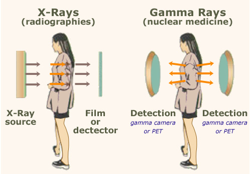
In traditional radiology, one measures the relative absorption of X-Rays passing through the body. In nuclear imagery, a handful of radioactive atoms (carefully chosen to latch on to the relevant organ) are injected into the body, and the gamma rays they emit from inside are detected to measure the concentration of radioisotope in the organ in question.
© BER
Scintigraphy scans involve the injection of a radiopharmaceutical product into the body – a radioactive source which will attach itself to an organ whose functions are of interest. It is for this reason that this field is referred to as ‘functional imagery’.
During an X-ray scan, detectors are set up to measure the number of X rays which pass through the patient’s body. In a scintigraphy scan, detectors are set up to count the number of rays emitted from the source located inside the patient. These measurements are carried out by sensitive gamma-cameras or PET-scanners, which are connected to powerful 3D imaging software to provide accurate inside maps of the patient’s body.
Scintigraphy scans today can be carried out on the lungs, the thyroid gland, the skeleton, the heart, the kidneys and even the brain. The procedures surrounding these exams differ depending on which organ is to be scanned, though the choices made as to the nature of the radiopharmaceutical product and the means of ingestion are also taken into account. When it comes to a cardiac scintigraphy, for instance, it is important to see the heart at two stages: at rest, and while pumping blood around the body. For this reason two separate scintigraphic scans need to be carried out.
Thanks to advances in detection and the possibilities offered by computers, simple scintigraphies are now giving way to SPECT (Single Photon Emission Computed Tomography). A tomoscintigraphy consists of a three-dimensional representation of a part of the body, from cross-sections, themselves obtained by the combination of planar scintigraphic images taken from various angles. The major applications of these tomoscintigraphies are the exploration of the heart, brain, lungs and skeleton.
These sophisticated Tomoscintigraphies are increasingly associated with scanners (TomoDensitoMétrie or TDM). This still rare association improves, for instance, the exploration sof the heart muscle function and of numerous bone lesions.
Example : Bone scintigraphies
Example : Cardiac scintigraphies
Other articles on the subject « Scintigraphic examinations »
Scintigraphic Diagnostics
A tool for non-aggressive analyses of biological functions Diseases are biological processes and [...]
Indications for Scintigraphy
When to Use Scintigraphy? The nuclear medicine physician must choose the appropriate marker produ[...]
Bone Scintigraphies
Principle and implementation of bone scintigraphies Bone scintigraphy is based on the fixation in[...]
Cardiac scintigraphies
Investigations of the cardiac muscle functionning MYOCARDIAL SCINTIGRAPHY BY PERFUSION Principle [...]