Low doses in diagnostics, strong and local with therapies
Radiation exposures in the medical field concern primarily patients who undergo examinations or treatment. This is the only case where exposures are delivered for the benefit of exposed persons.
The exposures differs strongly in the case of radiology or nuclear imaging diagnostics and the case of external or internal radiotherapies. In the first case, it is necessary to deliver the minimum dose in order to obtain a relevant diagnosis. In the second case, it is necessary to deliver the dose necessary to sterilize the tumour while preserving as much as possible the healthy tissues in the vicinity.
Medical exposures are not subject to limitations, unlike other sources of radiation due to man. Limitations would lead to the abandonment of valuable diagnoses for the patient. Radiation protection in this field is based on the principles of justification and optimization, i.e. to limit the use of radiation to what is necessary and when it is justified. In diagnostics, the constant improvement of the detectors sensitivity makes it possible to reduce in the same proportion the doses for the same result.
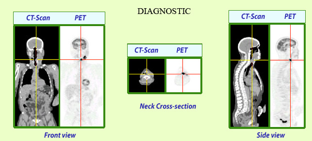
Exposition in the case of a combined PET Scan
The PET-SCAN examination has become a very effective tool for cancer screening and localization. This examination, which combines the diagnosis of a scanner (CT-Scan) and that of nuclear imaging (PET), exposes the patients to a dose of 10 to 20 milliSieverts, high for a diagnosis but justified by the importance of its result.
© IN2P3
In the case of nuclear or classical medicine radiodiagnostics, the doses of exposures generally considered are « whole body » effective doses. Low doses in conventional examinations such as a dental or pulmonary radiographies amount to a fraction of a millisievert. Exposures are much higher – several millisieverts – with much more extensive diagnostics, now commons, such as scanners (CT-Scans). Their nuclear medicine equivalents are scintigraphies (SPECT) and positron emission tomography (PET). An examination that combines scanner and PET becomes now frequently used (TEP-Scan) for cancers screening.
In the case of therapies, the irradiations are much more intense in order to destroy malignant cells. The irradiation should be concentrated as much as possible on the tumour and avoid the healthy cells in the vicinity. The doses applied to the tumours are of the order of tens of Grays.
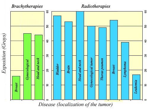
Local doses in therapies
In the case of radiation therapies, the exposures are local doses expressed in Grays. Doses are important because they are intended to destroy tumors. They are similar, whether it is nuclear medicine like brachytherapy on the left, or whether it is radiotherapy based on rays (right).
© IN2P3
Exposures to radiation in the vicinity of medical facilities concern also health professionals (doctors, radiophysicists, electroradiologists, nurses, …) who are involved daily in the use of ionizing radiation. In the early days of X-rays radiology, when one was not aware of their dangers, radiologists were the victims of a lack of protection. The diseases of radiologists belong now to a distant past. Today radiation protection measures have been put in place. In France, installations are subject to the strict regulations and controls of the Nuclear Safety Authority (ASN). Medical staff (149,000 persons in 2005) involved in the uses of ionizing radiation are subject to dosimetric monitoring of their exposures.
Finally, the general population should not exposed to waste or effluents from nuclear medicine services. These services use storage tanks for their liquid radioactive waste and effluents. In these tanks decline of radioactive products is generally rapid.
Articles on the subject « Expositions in Medicine »
Radiation doses in diagnostics
From simple radiology to PET scans and scintigraphy Scintigraphy scans are by far the most common[...]
Doses (X-Rays, CT scans)
Exposures to radiations in diagnostic radiology The vast majority of diagnoses based on radiation[...]
Doses (nuclear diagnostics)
Nuclear medicine : doses in nuclear diagnostics Nuclear medicine examinations are performed only [...]
Doses with therapies
Radio and nuclear therapies: Doses are high but local Genuine nuclear therapies (brachytherapy, p[...]
Doses in Nuclear therapies
High local doses In nuclear therapy, the doses delivered to tumours amount to few tens of Grays. [...]