Fruitful combination of two medical imaging techniques
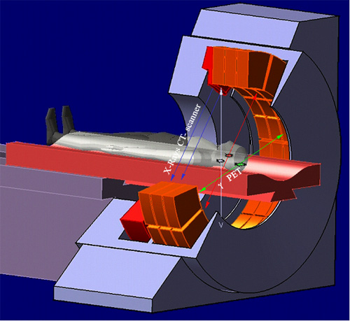
Diagram of a PET camera coupled with a scanner (CT)
Artist’s spatail view of PET camera combined with a scanner. Thanks to the annular arrangement of detectors, PET data are taken simultaneously at all angles.
© D.Steyaert/IN2P3
One of the latest technological advances in medical imaging for cancer screening is to combine « computed tomography » to « positron emission tomography”. This gives information and a diagnosis not available with other imaging techniques such as stand alone computed tomography (CT) or Magnetic Resonance Imaging (MRI).
An X-ray scanner is added to the most recent PET cameras (cameras PET / CT). The images produced by scanner are very precise from an anatomical point of view but provide little information on cell metabolism, that is to say, the changes within themselves of substances they contain. In contrast, PET images that are sensitive to this metabolism are called functional because they provide information on its working. But these images are less sharp and less precise from an anatomical point of view.
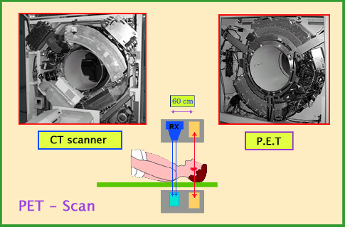
Principle of PET-CT
This diagram shows the principle of combining a scanner and a PET camera. The scanner and camera surround the bed where the patient is and move along the bed (axial movement). The PET camera detects annihilation photons from the radiopharmaceutical product, whereas X-rays from the scanner provides anatomical image of the body part examined.
© A.Aurengo/ Hôpital Pitié-Salpêtrière (Image Siemens)
In a hybrid device the anatomical images from the CT scan are merged with functional images made using the PET module. The combination of these two images can precisely locate an injury or malfunction. This will optimize a protocol during radiotherapy, make an operation more efficient, avoiding unnecessary surgical procedures or reduce the use of invasive procedures such as biopsies.
The scanner and the PET camera are located side by side in order to perform both tests one after another. As the patient does not move between the two examinations, the merging of two images is of excellent quality. Moving along the bed exam allow to cover the entire body.
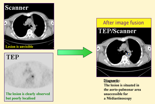
Combining PET-Scan(CT) data
The combination of a PET image and a CT image makes it possible to choose an appropriate procedure for a given case. The PET image for this patient shows the existence of a lesion in the mediastinum, the space between the two lungs. The scanner image locates the lesion exactly, which appears to be located in the aorto-pulmonary region inaccessible for a mediastinoscopy (invasive examination, requiring opening). There is a improvement in patient diagnostic
© General Electric/Medical systems
Due to the speed of the scan, less than 30 minutes for a global exploration, the results can be obtained in less than an hour compared to a day with previous technologies that require distinct examinations. It allows a more reliable and accurate diagnosis in cases of suspected cancer and for patients already sick.
This type of premium device, which combines radiology and nuclear medicine, is still rare in France. One of the first such device was unveiled in the Paris region in late 2002, in the center René Huguenin at Saint-Cloud. Since then, many PET / CT have been installed or are being installed. This screening device is in high demand. In 2014, a petition was circulating in the french West Indies island of Guadeloupe, asking to equip the island with a PET-scan, since the nearby sister Martinique island was already equiped … The problem has been resolved since.
RETURN : Positron Electron Tomography
Other articles on the subject « Nuclear Imaging »
Gamma Cameras
The most widespread of nuclear medical diagnostics Gamma cameras or scintillation cameras are pie[...]
Gamma-camera : principles
Detect gamma rays and reconstruct their line of flight The emission of a single gamma ray is a ve[...]
Positron Emission Tomography
PET : an instrument for early cancers detections Positron emission tomography, or PET, is a techn[...]
PET : Principles
Simultaneous detection of two annihilation gamma photons Positron Emission Tomography is an imagi[...]
Nuclear Imaging History
From radioactive indicators to CT, scintigraphy and PET … The name of Georg de Hevesy is as[...]