Gamma and PET cameras: powerful tools for diagnostics
The use of radiation has revolutionized the field of medical imaging technology. The inner workings of the human body can now be visualized with greatly increased accuracy – making radioactivity scans among the most important tools in the diagnostician’s arsenal. The two most common pieces of apparatus used in this field are gamma cameras and PET scans (Positron Emission Tomography scans). Of the two, gamma cameras are by far the most widely used.
Nuclear imaging as well as medical imaging in general benefits from many recent spectacular technical advances. Increasingly sensitive detectors reduce expositions while improving the quality of diagnosis. The development of digital technology now offers the physician immediate access to current or past exam data. For example, in the case of cardiac scintigraphies, imaging techniques are able to reproduce in real time on a screen the heartbeat of a patient.
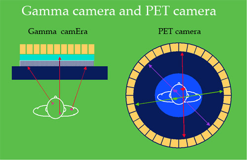
One or two gamma rays:
To obtain a clear picture with a gamma camera or a PET scan, a radioactive substance is inserted into the body then fixed to the organ in question. This insertion is often performed intravenously. Once the substance has been absorbed, a sensitive detection system is used to detect and analyse the emitted gamma rays. With a gamma camera, lead tubes are used to select the direction of photon emission. In PET, two photons simultaneously emitted back-to-back are detected : detectors are grouped in pairs, 180 degrees apart.
© IN2P3
Both types of camera were developed to detect photons travelling at very high energies – the only observable type of radiation that can pass through the human body. Gamma cameras, as the name suggests, are on the lookout for the appearance of gamma rays, whereas PET scans search for photons emerging from a matter-antimatter (electron-positron) collision. To obtain the desired images, the subject absorbs a radiopharmaceutical tracer specifically designed to fix itself to the affected area. Once in place, this tracer emits either photons or positrons, which can be measured and used to reconstruct an image of the organ in question.
Detectors and collimators allow to reconstruct approximate trajectories of the gamma photons. Even in the smallest samples of radioactive substances, millions of gamma rays are emitted every second. This large number of gamma photons allows to obtain finally highly accurate images. With such high numbers of trajectories, the calculations involved are so complex that they would be impossible without the help of modern computers.
Particularly efficient detectors are scintillators, currently used in gamma camera scans, which are called ‘scintigraphy’ scans. In scintigraphies, the tracers used are radioactive isotopes with half-lives; long enough to allow for their transportation to hospitals from the place of manufacture. These radioisotopes are often relatively heavy atoms (such as thallium or technetium or iodine) that can be attached by the relevant organs.
Gamma camera scintigraphy
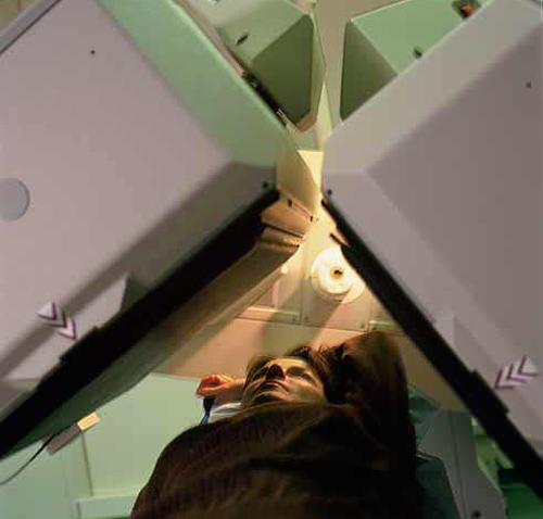
Gamma-camera detect emitted gamma photons : These cameras, the most common device in nuclear diagnostic , provides a functional image of an organ after administration to the patient of a specific tracer, usually injected intravenously, such as thallium-201 for the myocardium. Radioactive atoms emit photons captured by two planar detectors placed at 180 ° or 45 depending on the examination. The scintigraphies are routine clinical examinations that provide diagnosis, help to therapeutic decisions, disease evaluations and prognosis.
© CEA – L.MEDARD(SHFJ)
The most efficient positron emitter used in PET scanning is fluorine-18-desoxyglucose. Fluorine 18 is radioactive, and desoxyglucose is a compound that readily leaves fluorine 18 to attach itself to malignant tissue. As a result, this complicated compound is ideal for this kind of scanning.
The uses of PET are not just limited to the search for cancer – the technique can easily be applied to the search for specific chemicals (oxygen, carbon, nitrogen, etc.) which play an important role in the way living matter works. This makes scintigraphy scans immensely effective in analyzing the way living cells work. Some researchers have even used these techniques to film the workings of the metabolism.
The most significant contribution PET scans have made, however, is in the fight against cancer-related, cardiac and neurological problems. Such scans are able to reveal the abnormal working of tissue long before they can be picked up by scanners or regular MRI (magnetic resonance imaging) procedures.
Positron Emission Tomography (PET)
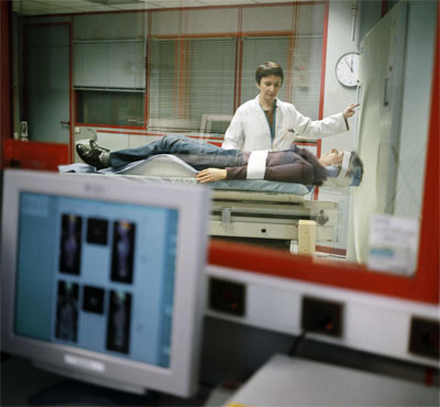
The radioactive tracers injected into the patient will emit positrons, which can be detected by a PET camera. This provides a reconstruction of the relevant organ and its metabolism; a highly practical and potentially life-saving procedure in brain scans, heart scans and mainly the screening of cancers. The entire scan takes around 30 minutes in cardiology, but can last for several hours when the entire body is scanned – as is the case in the search for metastasis.
© C.Boulze/CEA
The comparatively short life-spans of the radioactive elements involved make many of these techniques particularly expensive. The added cost, however, is compensated by the increased sensitivity and security they provide. Not only do they help cut down on unnecessary surgical procedures, they make it much easier to follow the progress of patients once they emerge from treatment.
Articles on the subject « Nuclear Imaging »
Gamma Cameras
The most widespread of nuclear medical diagnostics Gamma cameras or scintillation cameras are pie[...]
Gamma-camera : principles
Detect gamma rays and reconstruct their line of flight The emission of a single gamma ray is a ve[...]
Positron Emission Tomography
PET : an instrument for early cancers detections Positron emission tomography, or PET, is a techn[...]
PET : Principles
Simultaneous detection of two annihilation gamma photons Positron Emission Tomography is an imagi[...]
PET Scan
Fruitful combination of two medical imaging techniques One of the latest technological advances i[...]
Nuclear Imaging History
From radioactive indicators to CT, scintigraphy and PET … The name of Georg de Hevesy is as[...]