The most widespread of nuclear medical diagnostics
Gamma cameras or scintillation cameras are pieces of apparatus which allow radiologists to carry out ‘scintigraphy scans’, tests which provide detailed diagnoses about the functioning of the thyroid, the heart, the lungs and many other parts of the body. Scintigraphy scans get their name from the ability of some crystals (such as sodium iodide) to scintillate (in other words, emit sparks) when exposed to radiation.
Photomultipliers for gamma detection are now being replaced less bulky, more efficient and more accurate silicon detectors. The image resolution is improved, exposures for the same examination divided by 2 or sometimes by 5.
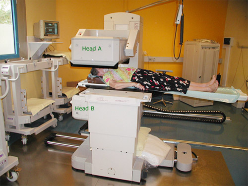
Double-headed gamma camera
This gamma camera is equipped with two heads able to detect the presence of radiation. The lower head is partly concealed under the bed.The whole apparatus can be moved along horizontally to obtain a full-body scintigraphy. By doubling the number of gamma rays used, a face scintigraphy can be performed at the same time as a back scintigraphy for the same amount of radioisotope ingested. By comparison with a PET scanner, a gamma camera requires much less equipment and is more easy to set up.
© CHU Avicenne
The procedure involves giving the patient a radiopharmaceutical molecule marked with a gamma-emitting radiosotope. Once the molecule fixed on the target organ or tissue, the highly penetrative emitted gamma rays easily escape from the body and leave their mark on the detection panels. The molecule which is followed around the body is carefully chosen with respect to the body part under examination. A very small quantity of radioactive isotope is all that is needed, as the detection systems are sensitive enough to register the decay of individual atoms.
Furthermore, scintigraphies taken at different angles is obtained by rotating the camera. Then, by combining these planar images, it is possible to reconstruct, thanks to computer science, tomographies,  3-dimensional spatial images. This technique based on gamma-cameras scintigraphies is called Single-photon emission computed tomography (SPECT). The basic information is typically presented as cross-sectional slices through the patient.
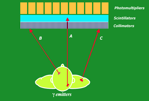
Principles of gamma-camera detection
In a gamma-camera, every decaying technetium radionuclei emits a gamma photon. After measuring the gamma impact position on the detector, one needs to know its direction to go back to its origin. A collimation is necessary. This collimation is obtained by lead channels selecting the photons travelling through. In the figure above, only the photon A reaching the scintillator will be detected by the photomultipliers, the photons B and C being absorbed by the lead.
© André Aurengo, Hôpital Pitié-Salpêtrière
As its name suggests, ‘gamma camera‘ detects scintillations produced by gamma rays emitted by a radioactive marker. The impact of these gamma rays on a sodium iodide crystal generates scintillations that are detected by photomultipliers. Once a large number of these scintillations have been observed, the radioactive emitters of these gamma rays can be located.
Scintillators and photomultipliers for the detection of gamma are now increasingly replaced by silicon detectors, less bulky, more efficient, more accurate. The resolution of the images is improved, while the exposures for an examination are divided by 2 or sometimes by 5.
Thanks to computer technology, complex calculations can be performed very quickly to convert the detected radiation into information that is useful for radiologists. The images, created in a fraction of a second, allow doctors to follow the spread of the radioisotope throughout a patient’s body in real time. This allows for highly detailed pictures of the heart contraction, or of the filtration of blood plasma in the kidneys. A gamma camera scintigraphy can also be used to form images of the skeleton, by injecting patients with a radioactive solution which attaches itself onto the bones. This is often how skeletal metastases are detected.
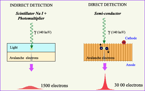
Two detection techniques
The oldest technique on the left is that of a scintillator crystal associated with a photomultiplier. The impact of a gamma generates a light signal, amplified through an avalanche of electrons by the photomultiplier.This technique is replaced today by the use of semiconductors (right). The gamma impact directly triggers a much larger avalanche of electrons.
© Source Université Paris-Sud
The scintillation camera was invented by the American physicist H.O. Anger in Berkeley in 1957. It has since revealed itself to be an irreplaceable tool in a wide range of different diagnoses. Unquestionably the preferred piece of equipment in the field of nuclear medicine, there were 14,000 of them in the world by 1996 and much more now.
NEXT : Gamma camera : principles
Other articles on the subject « Nuclear Imaging »
Gamma-camera : principles
Detect gamma rays and reconstruct their line of flight The emission of a single gamma ray is a ve[...]
Positron Emission Tomography
PET : an instrument for early cancers detections Positron emission tomography, or PET, is a techn[...]
PET : Principles
Simultaneous detection of two annihilation gamma photons Positron Emission Tomography is an imagi[...]
PET Scan
Fruitful combination of two medical imaging techniques One of the latest technological advances i[...]
Nuclear Imaging History
From radioactive indicators to CT, scintigraphy and PET … The name of Georg de Hevesy is as[...]