When to Use Scintigraphy?
The nuclear medicine physician must choose the appropriate marker product for the examination. The marker must associate with the desired part of the body in a highly specific manner.
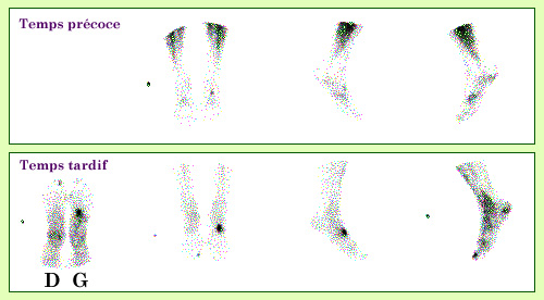
Bone Scintigraphy, a Diagnostic Tool We use nuclear diagnosis when it represents the only means of obtaining the desired information quickly and safely. This is seen in the case of this 34-year-old patient who suffered from persistent pain while walking and had normal X-rays. Diphosphonates labeled with technetium-99m have an elective affinity for bone tissue in remodeling (cancer metastases and areas of inflammation). A bone scintigraphy established that these disorders were due to a metal drawer falling on the left foot three months before the examination.
Source: A.Aurengo/Hôpital La Pitié Salpétrière
We can use:
- A simple radioactive atom, an unstable isotope of an element that exists naturally in the body. This is the case for several isotopes of iodine, which are known to concentrate in the thyroid gland, making them an excellent marker for this gland. We can also use atoms that are metabolized in the body in a similar way to a natural substance, if the latter does not have a radioactive isotope that emits usable radiation. For example, thallium-201 behaves like potassium, which is widely captured by muscles, particularly the heart muscle.
- A labeled molecule. It is essential to ensure that the labeling is stable during the various phases of normal human metabolism and that the degradation of the new molecule does not lead to the appearance of toxic components. For example, diphosphonates labeled with technetium-99m have an elective affinity for bone tissue in remodeling (areas of inflammation or cancer metastases). However, we can also link a radioactive atom to a hormone or an antibody.
- A set of cells, taken from the subject, kept alive, and labeled with the radioactive tracer, then reinjected into the patient to follow their behavior. For example, red blood cells labeled with technetium-99m allow us to visualize the cardiac cavities and calculate various important parameters in the study of cardiac dysfunction.
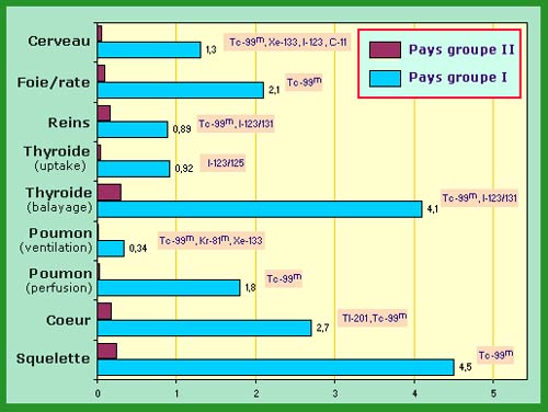
Frequency of Scintigraphies Annual frequency of scintigraphic examinations per 1000 inhabitants for countries classified according to the quality of their healthcare system. The first group includes countries like Canada, the United States, Japan, and European Community states; the second group includes Brazil, China, India, etc. Within a group, medical practices vary from one nation to another. The average of the results of several countries differs from the results of France alone.
Source: IN2P3 (Source: K.G.Gerber. Adapted from UNSCEAR 2000)
It is evident that all the manipulations required for the preparation of injections must be performed with great care, and strict controls are present at each stage.
Currently, almost all medical specialties can use radioisotopes, whether for diagnostic (imaging or biological assays) or therapeutic purposes. However, certain areas are particularly privileged, and there are even areas where nuclear medicine represents the only means of obtaining the desired information quickly and safely.
Among the main indications in our country, we can mention:
- First and foremost, bone and joint disorders (a little more than a third of scintigraphies performed in France), with a large number of searches for bone metastases (breast cancer, prostate cancer), but also many less severe pathologies (assessment of various pains and infections affecting bone tissue).
- Cardiovascular exploration (25%), grouping examinations of the myocardium (coronary pathology) and imaging of the cardiac cavities (isotopic ventriculography and ejection fraction calculation).
- Pulmonology, particularly searches for pulmonary embolisms (15%), the only area where scintigraphy is used in emergency and is a first-line examination that can orient diagnosis and adapted therapy.
- Thyroid, the first and largest indication of nuclear markers, now less explored in nuclear medicine (10-12% of examinations), given the highly performant ultrasound techniques also used in this field.
- Finally, a set of scintigraphies dedicated to renal, digestive, and neurological explorations.
There is also a vast domain of applications in biology that concerns all immuno-assays using antibody-antigen reactions labeled with radioisotopes. These techniques, although largely competed by advances in immunofluorescence, remain sometimes the reference (hormone assays, drug assays, various pathogen assays). In the field of hematology, radioisotopic techniques remain irreplaceable for the study of the functionality of different organs and blood cells involved in hemopathies.
Other articles on the subject « Scintigraphic examinations »
Nuclear Scintigraphies
A range of diagnoses for determining how the body works … The word ‘scintigraphy&lsqu[...]
Scintigraphic Diagnostics
A tool for non-aggressive analyses of biological functions Diseases are biological processes and [...]
Bone Scintigraphies
Principle and implementation of bone scintigraphies Bone scintigraphy is based on the fixation in[...]
Cardiac scintigraphies
Investigations of the cardiac muscle functionning MYOCARDIAL SCINTIGRAPHY BY PERFUSION Principle [...]