Detect gamma rays and reconstruct their line of flight
Gamma camera with parallel collimators
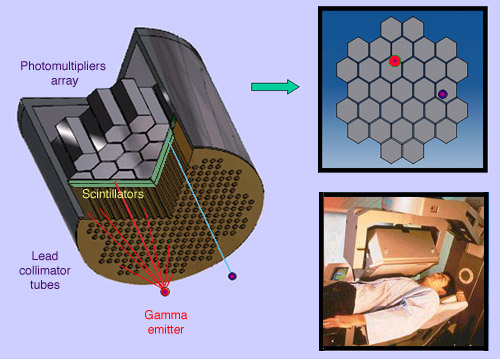
Principle of gamma detection using a collimator with parallel channels. Only gamma propagating along the channel axis reach the scintillator, where they deposit their energy, generating a brief flash of light. The scintillation is detected by an array of photomultiplier behind the scintillator back. Photomultiplier converts the light pulse into an electronic signal that is amplified by the camera electronics. The impact position and gamma energy are evaluated from the sthe photomultipliers data. The insert shows a gamma camera pointed at the chest of a patient.
© D.Steyaert/IN2P3
The emission of a single gamma ray is a very small-scale nuclear phenomenon. It is the role of the gamma-camera head to amplify this microscopic radiation into an electric signal that can be detected and measured. By exploiting a large number of readings of these electric signals, one can determine the map of the radioactive nuclei responsible for the emission of gamma rays.
The gamma-camera detection head consists of:
– a collimator
– a scintillating crystal
– an array of photomultiplier tubes
– an electronic system for detection and measurement of gamma energies and impacts
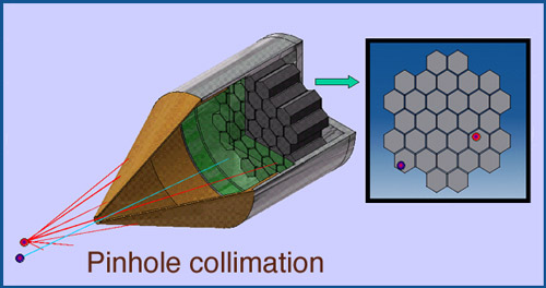
Gamma camera equipped with a pinhole collimator
For thyroid scans gamma cameras equipped with a pinhole collimator are used, also known as « sténopéic » . For a small gland like the thyroid this type of collimation is appropriate. The tip of the camera is directed toward the patient’s neck. Only gamma passing through the pin-hole, coming from the thyroid zone, will be recorded.
© D.Steyaert/IN2P3
The collimator is a thick plate of lead or tungsten riddled with a large number of very thin parallel channels. The gamma rays which can pass through it are those whose direction is perpendicular to the surface of the lead plate and the scintillating crystal. The channel axes point towards the body part under examination, and the lead or tungsten stops all gamma photons travelling at an oblique angle. Other collimators can be designed using different techniques: a pinhole collimator is used for thyroid scintigraphy scans, whereas fan-shaped collimators are used for imaging of the brain.
The detection element at the heart of a gamma camera is a large rectangular crystal of sodium iodide doped with thallium: NaI (Tl). The crystal has the ability to stop incoming gamma rays and convert part of the deposited energy into scintillations.
Behind the crystal, an array of small photomultipliers converts photons of light into electric signals. From the hits in a set of photomultipliers, one can determine the energy of the incoming gamma rays as well as the approximate position of their impacts on the crystal. The gamma rays whose energies do not fall within a certain range of the energy of the radioactive sample (a spectroscopic window) are discarded, and do not contribute to the final picture.
The gamma camera is positioned in such a way as to ensure that it selects the gamma photons being emitted by the organ under diagnostics.
A semi-conductors based gamma imager
Performances of scintigraphy and PET gamma-cameras benefit of major recent advances in detection techniques. For instance, a team of CEA physicists proposed in 2015 a gamma imager based on Cadmium-Zinc-Telluride semiconductor, much more accurate in measuring a gamma energy and position.
© Clés CEA N°200
The images quality depend on the accuracy of reconstruction. Conventional detection with scintillators associated to photomultiplier will be gradually replaced by more accurate detection systems. For instance the accuracy of the position of gamma impacts may improve from 3 mm to 0.3 mm with based semiconductors imagers.
Other articles on the subject « Nuclear Imaging »
Gamma Cameras
The most widespread of nuclear medical diagnostics Gamma cameras or scintillation cameras are pie[...]
Positron Emission Tomography
PET : an instrument for early cancers detections Positron emission tomography, or PET, is a techn[...]
PET : Principles
Simultaneous detection of two annihilation gamma photons Positron Emission Tomography is an imagi[...]
PET Scan
Fruitful combination of two medical imaging techniques One of the latest technological advances i[...]
Nuclear Imaging History
From radioactive indicators to CT, scintigraphy and PET … The name of Georg de Hevesy is as[...]