Doses (nuclear diagnostics)
Nuclear medicine : doses in nuclear diagnostics
Nuclear medicine examinations are performed only if they are of benefit compared to conventional diagnostic. As radiologists, nuclear physicians should assess the importance of the exposure resulting from such an examination and how this exposure varies depending on its circumstances. This is to minimize the risk.
Irradiating power
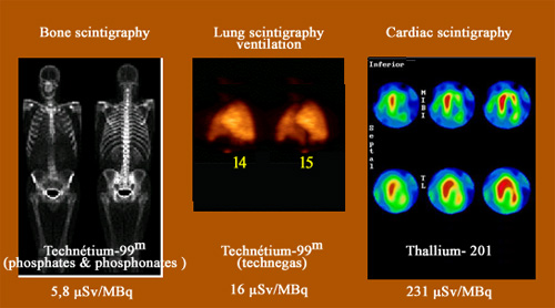
The irradiating power of an examination is defined as the dose resulting from the injection of one million Becquerels (MBq). If one express the dose in micro-sievert (µSv) (millionths of a sievert), the irradiating power is measured in microsievert / MBq. This irradiating power varies in large proportions from one type of scan to another. Bone scans are almost not irradiating (5.8 microsievert / MBq), when heart scans are much more irradiating (231 microsievert / MBq).
© IN2P3/ Source ICRP : publication 80).
The exposure levels are proportional to the activity of the radioactive tracer at the time of injection and depend on the length of stay in the body (until its physical or biological removal). They obviously depend on the selected tracer. Activity at the time of injection is evaluated in million of Becquerels (MBq or mega-Becquerels). However, in France many practitioners still rely on traditional millicuries, unit still widely used (1 mCi equals 37 MBq).
The injected activities vary greatly depending on the exam, as for example from 1 mCi for kidney scintigraphy with iodine-123 to about 27 mCi (1000 MBq) for cardiac scintigraphy with technetium.
The exposure is naturally proportional to the injected activity but also on the irradiating power of the radioactive substance. This irradiating character depends on the nature of radiation, how long it stays in the body, and how the radioactive isotope is distributed in the patient body. It varies widely.
Scintigraphy by far the most irradiative is the heart muscle with thallium-201. The resulting dose injection of 150 MBq of thallium is 37 millisieverts (mSv) which is equivalent to 16 years of natural irradiation! If one believes in the validity of ICRP linear relation without threshold to evaluate the risk of cancer at this dose, the risk of cancer is about 1 in 500. This potential risk of cancer that would make ten or more years to be declared must be weighed against the risk that has lead to the examination. The patient is at risk of a fatal outcome in the short term if a severe heart defect – like a clogged artery – is not detected and treated.
The positron emission tomography (PET) is still rare compared to scintigraphy. Most of these tests use as a marker 18F-FDG whose irradiating character is estimated by the ICRP – for an adult – to 0,019 mSv / MBq. For a standard PET (injected activity of 10 millicuries or 370 MBq), the resulting dose is 7 mSv, about the same as for a scanner. If tomography is performed in conjunction with a scanner (PET-SCAN) the dose from the scanner must be added.
Other articles on the subject « Expositions in Medicine »
Radiation doses in diagnostics
From simple radiology to PET scans and scintigraphy Scintigraphy scans are by far the most common[...]
Doses (X-Rays, CT scans)
Exposures to radiations in diagnostic radiology The vast majority of diagnoses based on radiation[...]
Doses with therapies
Radio and nuclear therapies: Doses are high but local Genuine nuclear therapies (brachytherapy, p[...]
Doses in Nuclear therapies
High local doses In nuclear therapy, the doses delivered to tumours amount to few tens of Grays. [...]