Scintigraphic Diagnostics
A tool for non-aggressive analyses of biological functions
Diseases are biological processes and Nuclear Medicine offer the advantage of providing images of these biological processes. Most of the radiotracers are part of a biological process – such as renewal of minerals in the bones, potassium transport in cardiac muscle, or glucose (sugar) metabolism in various organs or tumors. Because it provides images of a biological process (physiology), Nuclear Medicine differs from other imaging techniques like X-ray scanners, nuclear magnetic resonance, and ultrasound that visualize especially the structures and forms (anatomy).
In France, Nuclear Medicine is currently a medical specialty of its own, accessible to medical students only at the level of the « internat », the final stage of the French medical curriculum. It differs from other specialties by compulsory formation on nuclear physics and techniques held at INSTN, Saclay. It is defined as encompassing all the techniques using radioelements in humans, either for diagnostic or therapeutic purpose.
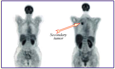
Cancer screening with PET
Nuclear medicine examinations allow to see what conventional tests cannot diagnose. The figure shows two « full body scans » obtained on two different patients by positron emission tomography (PET). Left image is normal: the right image is that of a patient with a lung tumor developing after a primary breast cancer. The scan shows an increased consumption of F-18 FDG in the tumor, the developing tumors using more sugar (glucose) than the surrounding normal tissues.
© DOE/BER
Features and techniques
Three features should be stressed about the use of the imaging techniques applied in Nuclear Medicine for diagnostic (scintigraphy and PET).
– it is relatively harmless because radioactivity amounts introduced into the human body are minimal. Most scans deliver doses of radiation in the range of simple chest radiography. Many studies now tend to further reduce doses to obtain identical information, or even more precise ones;
– it is not aggressive, because except in very particular cases, a single intravenous injection is enough, the product diffusing into the body and focusing more or less in some tissues as a function of known affinity;
– it provides a functional imaging, meaning that it provides information not only on the morphology of the studied organ, but also and especially on its operation mode. For example, a thyroid scan, giving a fairly accurate picture of the distribution of iodine in the thyroid may be complemented by a study over time that will judge of the normality or not, of the fixing and removing process of iodine. This is related to the peculiarity (very valuable in this case) of the radioactive body to behave exactly as the stable isotope and to have the same metabolism, which can be followed by studying emitted radiations.
Nuclear medicine practicians use their knowledge in biology and pharmacokinetics to determine the best-suited marker for the diagnosis sought. Later on, during the examination, the required handlings for the preparation of injections should be done with utmost care and strict controls accompanying each step of the procedure.
Learn more :
Other articles on the subject « Scintigraphic examinations »
Nuclear Scintigraphies
A range of diagnoses for determining how the body works … The word ‘scintigraphy&lsqu[...]
Indications for Scintigraphy
When to Use Scintigraphy? The nuclear medicine physician must choose the appropriate marker produ[...]
Bone Scintigraphies
Principle and implementation of bone scintigraphies Bone scintigraphy is based on the fixation in[...]
Cardiac scintigraphies
Investigations of the cardiac muscle functionning MYOCARDIAL SCINTIGRAPHY BY PERFUSION Principle [...]