An ultra-sensitive analysis of surface composition
Editorial review 2026
The analysis technique mostly used the AGLAE the accelerator is based on the detection of X-rays. The PIXE (Particle Induced X-ray Emission) technique uses X-ray emission induced by accelerated charged particles that interact with the atoms of the materials encountered in the same way as radiation emitted by radioactive processes. When the particle pull out one of his innermost electrons of an atom, the atom reacts by emitting an X-ray of characteristic energy.
These X-rays are able to get out of the material analyzed. They are produced in large numbers, they can be detected and their energy measured. From the energy spectrum of these X-rays photons, the fundamental composition of the sample can be deduced.
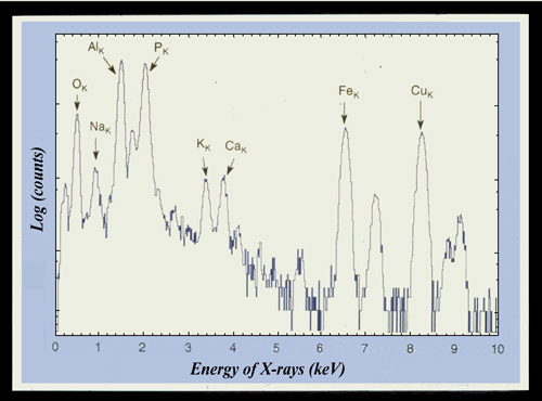
A PIXE spectrum : characters of a coptic manuscript
The energy distribution of X-rays emitted by Coptic manuscript characters shows the presence of traces of iron. These traces of iron are characteristic of the « metallo-gallic » nature of the ink used. The galls (oak parasite) macerated in vitriol, was used to make black ink.
© LRMF
The method is similar in principle to X ray fluorescence used in industry. But while the radiation used in X-ray fluorescence (gamma or X rays) penetrates deeply into the material, it is the surface of the sample that is analyzed with PIXE, since the ions coming from the accelerator stop after few microns.
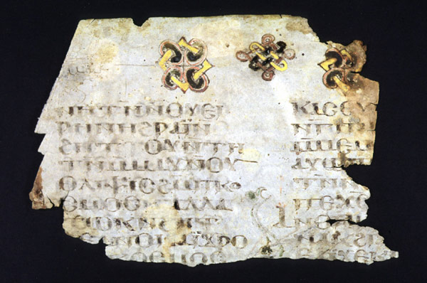
Coptic manuscript
The surface of the analyzed object impacted by the particles delivered by the accelerator AGLAE is extremely small: a fraction of a millimeter square. The extreme precision of the particle beam allows to point to each of the characters of this Coptic manuscript of the thirteenth century and to analyze by the PIXE technique the nature of the inks used.
© LRMF
The advantages of the PIXE technique are numerous. It detects all elements heavier than sodium. It is fast: irradiation of one minute is sufficient. It is sensitive: allowing to measure levels of a few parts per million. It does not damage the sample, which is great for valuables (jewellery, paintings, paper…).
Finally, this technique is local: the beam of projectiles, the diameter of which can be chosen between a few millimetres and one micron, can study such details as a weld in a piece of jewellery or an inlay.
The accelerated particles penetrate little in the target and only probe the surface. Thus, two million electron volts protons allow analyzing a piece of silver of 10 microns thickness. For potteries, the path of protons does not exceed a few tens of micron.
This technique has indeed an essential feature in this field of investigation : It is non destructive. A breakthrough has been to develop an experimental device allowing direct analysis without necessitating a sample of the object. This is achieved by extracting the beam in air through an exit window ultra-thin, down to 0.1 microns thick.
Two other analysis techniques are also used, sometimes in combination with the PIXE technique : the emission of gamma induced by nuclear reactions (PIGE method) ; The « Rutherford backscattering (RBS) which identifies the nature of nuclei from beam particles which bounce back.
The applications of Ion Beam Analysis to art and archeology back almost to the beginnings of the development of PIXE technique, with study of ceramic fragments and obsidian tools. They were then quickly extended to all cultural heritage materials.